Molecular Cytogenetics:Target and Probe-DNA Denaturation
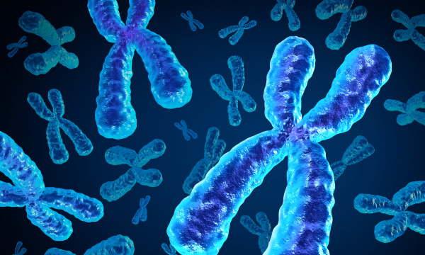
Molecular Cytogenetics integrates the fields of molecular biology and cytogenetics to examine the chromosomal structures in order to distinguish between healthy and cancerous cells.
1) the historical perspective of its development from cytogenetics, 2) technical issues, 3) accessible probe sets, and 4) variants and uses of the fundamental fluorescence in situ hybridization (FISH) approach The term "cytogenetics" and/or the use of whole-genome oriented molecular genetic methods are being replaced by the term "cytogenetics" in the current zeitgeist. So, even though " cytogenetics" is clearly a "cytogenetic approach," a change from "Molecular Cytogenetics" to " cytogenetics" is not justified by any means, a few remarks on this point are required to understand why a change from " cytogenetics" to "MC" is not justified by any means.
Following that, technical features of fluorescence in situ hybridization are discussed, as well as how cytogenetics and Molecular Cytogenetics were developed (FISH). The latter includes the methodology, as well as probe and target DNA types and probe sets. The primary section focuses on instances of current FISH-applications, emphasizing the approach's unique capabilities, such as the ability to analyze individual cells and even individual chromosomes.
Besides second and third-generation sequencing techniques, many versions of FISH can be utilized to retrieve information about genomes from (nearly) base pair to the entire genomic level. Molecular combing, chromosome orientation-FISH (CO-FISH), telomere-FISH, parental origin determination FISH (POD-FISH), FISH to resolve nuclear architecture, multicolor-FISH (mFISH) approaches, among others used in chromoanagenesis studies, Comet-FISH, and CRISPR-mediated FISH-applications are among the FISH variations highlighted here. Overall, Molecular Cytogenetics is far from obsolete, and it is used in cutting-edge diagnosis.
Target and Probe-DNA Denaturation
• Incubation of target- and probe-DNA in a hybridization solution at 37°C for several hours (with or without background-blocking repetitive DNA sequences);
• Using appropriate buffers to remove any extraneous probe-DNA;
• If necessary, use a fluorophore-labeled antibody to detect the hapten attached to probe-DNA; if probe-DNA is already fluorescence-labeled, use antifade-solution and a coverslip;
• A fluorescence microscope examination.


Comments
Post a Comment