Amniotic Membrane Transplant: Surgical Technique and Procedures
 |
| Amniotic Membrane |
Surgical Technique
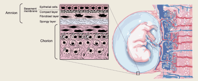
The Amniotic Membrane, or amnion, is the deepest layer of the placenta and comprises a thick storm cellar film and an internal stromal lattice. Amniotic film transplantation has been utilized as a join or as a dressing in various careful subspecialties. In the area of ophthalmology, it is utilized comprehensively 1) to reproduce the visual surface after different techniques; 2) as a join for visual surface melts; and 3) as gauze to advance mending in instances of diligent epithelial deformities or visual surface aggravation. These signs utilize amniotic film's capacity to advance recuperating.
Mechanism of Action
The cellar film part of the amniotic layer is comparable in synthesis to the conjunctiva. Thus, the current hypothesis proposes that the amniotic layer expands support for epithelial cells, limbal immature microorganisms, and corneal transient enhancing cells. Clonogenicity is kept up with which advances both flagon and non-challis cell separation while barring incendiary cells and their protease exercises. Moreover, the stromal face of amniotic film smothers myofibroblast separation of typical fibroblasts to diminish scar and vascular arrangement. This activity helps with mending for conjunctival reproduction, epithelial deformities, and stromal ulceration.
Pterygium Excision
After careful expulsion of pterygium, a conjunctival imperfection remains. This imperfection could be left alone to mend by auxiliary aim, stitched straightforwardly by means of essential conclusion, united with a conjunctival autograft, or joined with an amniotic layer. With intraoperative steroid infusion into the encompassing tissue imperfection, amniotic layer transplantation has an equivalent repeat rate to conjunctival autograft. The use of mitomycin-C doesn't further decrease the repeat rate as indicated by certain examinations. There are many clashing reports in regards to the pace of postoperative pterygium repeat after the utilization of the amniotic layer. Amniotic Membrane could be viewed as when the size of the employable deformity would be dangerous for the direct conclusion, or when a conjunctival autograft can't be acquired due to scarring or the presence of glaucoma gadgets.
Limbal Stem Cell Deficiency or Persistent Epithelial Defects
Amniotic Membrane can be utilized in instances of fractional and absolute limbal undifferentiated organism insufficiency. In instances of complete limbal immature microorganism misfortune, AMT alone won't do the trick and should be utilized related to allogeneic undeveloped cell transplantation. For instances of halfway limbal undifferentiated organism misfortune, and furthermore, on account of diligent epithelial imperfections, an amniotic layer has been displayed to upgrade epithelialization and further develop a vision with and without allogeneic limbal cell transplantation.
Procedure
Many reports in the writing delineate the utilization of the human amniotic layer collected from the placenta at the hour of the cesarean area and protected until use on the visual surface. The Cryopreserved amniotic layer is accessible and generally utilized, and it holds the histological and morphological properties of new tissue. The amniotic layer can be precisely connected to the visual surface by absorbable or non-absorbable stitches. Organic tissue glues have additionally been utilized to append Amniotic Membrane to the visual surface.


Comments
Post a Comment